下載 APKPure App
可在安卓獲取HIV Virus Structure in 3D VR的歷史版本
HIV(人類immunodefficiencyvirus)這裡無害的,免費為您去發現!
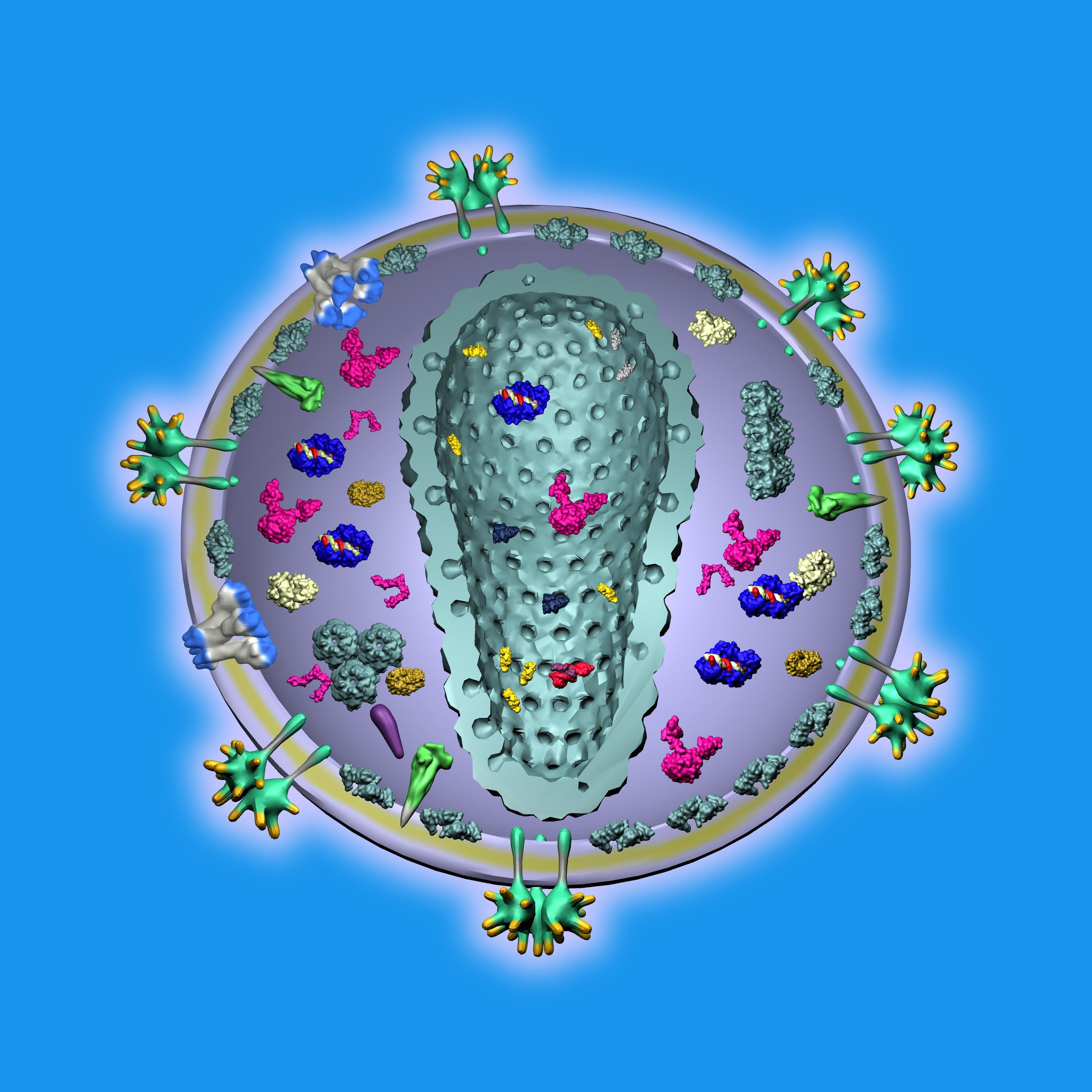
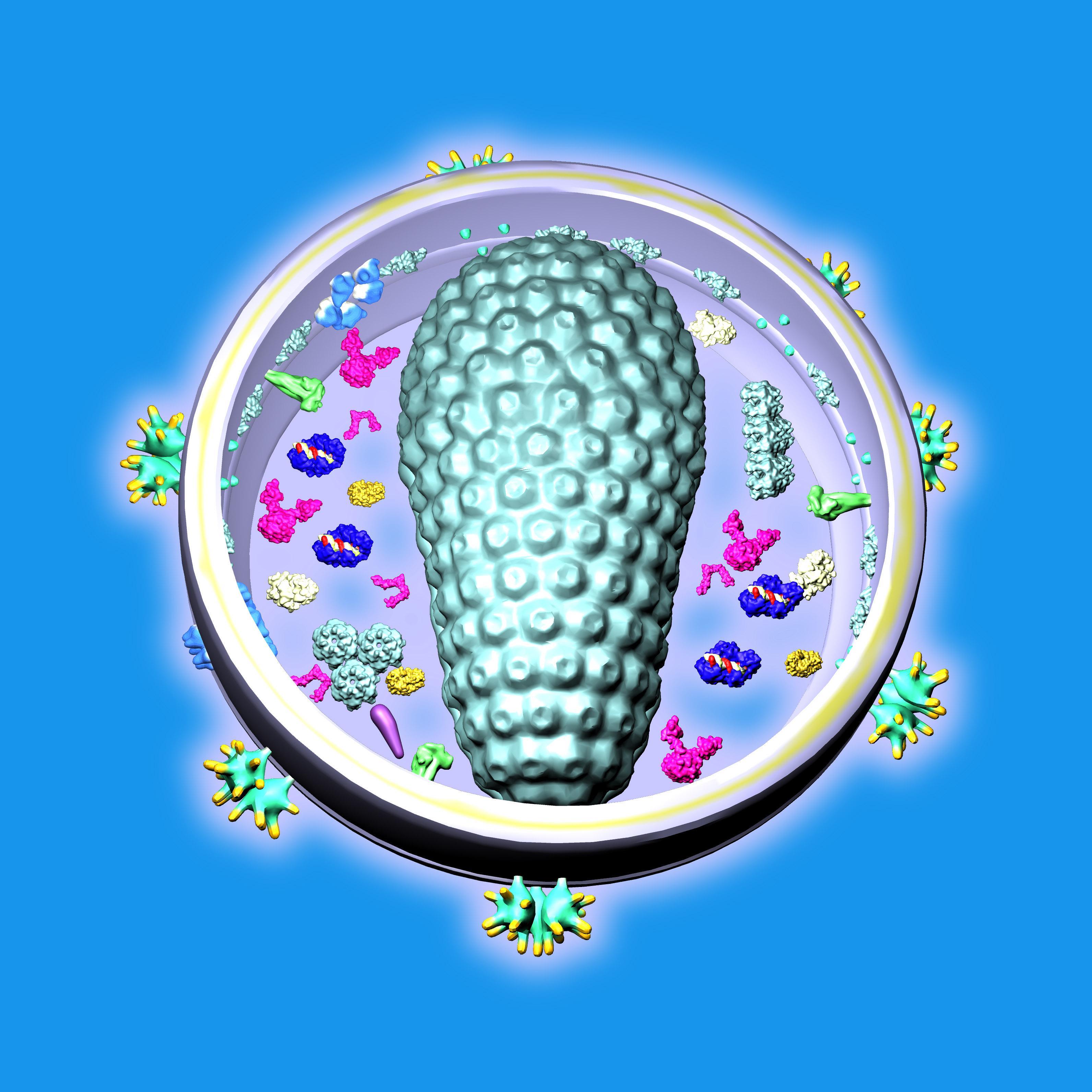
HIV (Human immunodefficiencyvirus) is composed two copies of the single stranded RNA, 5 types of stuctural proteins, 3 types of viral enzymes, and 7 types of accessory proteins along with some proteins taken from the host cell. Its central capsule of the oval shape is surrounded by the round vesicle made of the lipid bilayer (=elementary cell membrane).
Structural proteins:
The most prominent are the envelope proteins gp120 and gp41 that "grow" like trees from the outer lipid bilayer membrane. They bind the HIV vesicle to the receptors on the membrane of the host cell. They bear a lot of carbohydrates on the outer sides that make them difficult to be recognized by the antibodies.
Matrix proteins form the trimers and are inserted into the inner side of the lipid bilayer membrane and form the inner coating of the outer viral vesicle.
Capsid proteins form the hexamers and constitute the coat of the inner oval core of the HIV virus which surrounds the viral RNA. Some of them are located also freely in the intermediary space between the core and the outer membrane vesicle. Capsid proteins should not be confused with the nucleocapsid proteins. Nucleocapsid proteins protect the viral RNA.
Viral enzymes:
Reverse transcriptase produces DNA from the viral RNA template. This DNA then coded in the host cell for the new copies of the HIV virus. After the DNA is built, the original RNA is destroyed. Some HIV drugs work by blocking this enzyme.
Integrase inserts the DNA (that has been produced by the reverse transcriptase) into the host genome. So the viral DNA can live inactive for a long time and is very difficult to identify and fight.
HIV protease enables the formation of the HIV particles. Originally the building blocks for HIV have the form of long protein chains which have to be segmented into the final proteins. This task is performed by the HIV protease.
Accessory proteins:
Viral protein u helps the newly formed viral vesicle to escape from the host cell by inhibiting the attractive force between the viral proteins and the cell receptors. As its form suggests, this protein also functions as the membrane channel. Viral infectivity factor binds to the defense proteins of the cell.
Viral protein r help the viral genome to reach the nucleus of the cell.
P6 helps in the formation of the new viruses. Its form has not yet been determined.
Negative regulatory factor is responsible for the weakened response of the cell. It inhibits the formation of several proteins playing a role in the defense of the cell.
Regulator of virion protein binds to the viral RNA and governs its segmenting and travelling through the cell.
Trans activator of transcription binds to the viral RNA.
Last updated on 2016年06月16日
Minor bug fixes and improvements. Install or update to the newest version to check it out!


HIV Virus Structure in 3D VR
1.2.1 by Bioanim
2016年06月16日